Clinical Example: Percutaneous fixation of proximal fracture of the proximal phalanx
|
Fractures of the proximal part of the
proximal phalanx are often unstable and angulate
dorsally. Longitudinal
percutaneous fixation may not provide adequate purchase
on the proximal
fracture fragment. These images show a method
of increasing stability of percutaneous fixation in this
situation by placing additional pins into the proximal
fracture
fragment and incorporating these into an external
fixation framework. The metacarpophalangeal joint
capsule is entered with the pins, but the pins do not
engage the metacarpal head. |
| Click on each image for a larger picture |
|
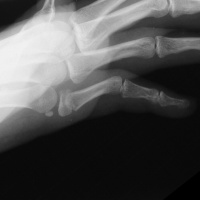
Case 1.
A malaligned three
week old fracture. Notice the
typical zig zag collapse pattern resulting in flexion
of the proximal interphalangeal
joint to the same degree as the fracture angulation. |
| Intraoperative
fluoroscopy. |
| Manipulation and closed reduction: |
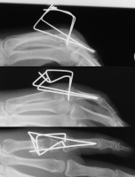
| Percutaneous pins placed across the fracture line and also dorsal to palmar into the proximal fracture fragment. |
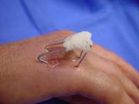
| The pins were left protruding through the skin, and were bent toward each other to form a common zone of overlap which was glued together with aquaplast to form an external fixation construct. |
| With this
technique, the hand is immobilized in a protective
position splint for four weeks. Immediately before pin
removal. |
|
Case 2.
Unstable dorsal and laterally angulated proximal
phalanx fracture in an elderly osteopenic woman. |
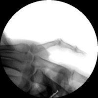
| Closed reduction and insertion of interfragmentary and proximal fracture fragment pins. |
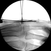
| Pins were bent to
overlap and then bonded with heated thermoplastic
splint material. |
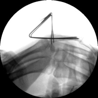
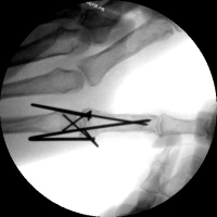
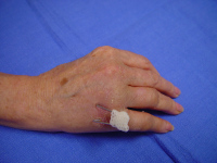
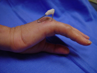
| Four weeks postop,
immediately prior to pin removal in the office. |
|
Search for...
percutaneous fixation phalanx proximal phalanx fracture |
Case Examples Index Page | e-Hand home |