Clinical Example: Enchondromas of the distal phalanx
| Enchondromas are benign intraosseous tumors which present with enlargement, pathologic fracture, or occasionally pain from incipient fracture. They are effectively treated with curettage, with or without bone grafting, and have a low recurrence rate. |
| Click on each image for a larger picture |
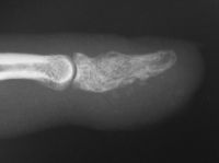
| Case 1. This patient presented with pain with thumb pinch and a sense of fullness in the thumb pulp. Plain radiographs demonstrate an expansile, geographic, radiolucent, juxtaarticular intraosseous mass, typical for an enchondroma. There is circumferential cortical thinning and possible cortical breaks. |
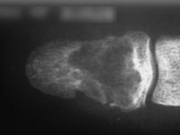
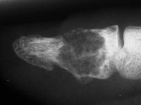
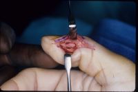
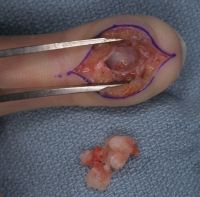
| Because the palmar cortex appeared to be the strongest remainig area, a midlateral approach was chosen over a midline volar longitudinal incision. |
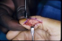
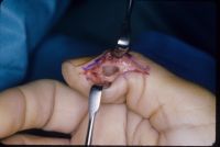
| A corticocancellous strut bone graft was placed in the defect to stabilize the weakened bone. |
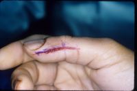
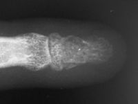
| Late result. |
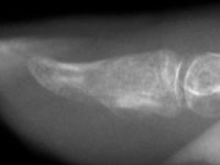
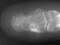
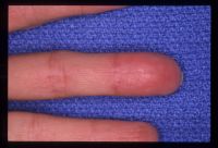
| Case 2. This patient presented months after having "injured" her middle fingertip, feeling that the finger was not normal. Xrays were consistent with an enchondroma and probable dorsal angulation from a healed pathologic fracture. |
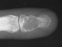
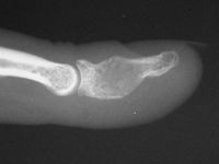
| This was excised through a midline palmar incision. The cavity was debrided with a high speed burr, then packed with cancellous bone. |
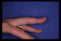
| Final result. |

|
Search for... enchondroma distal phalanx tumor
|
Case Examples Index Page | e-Hand home |