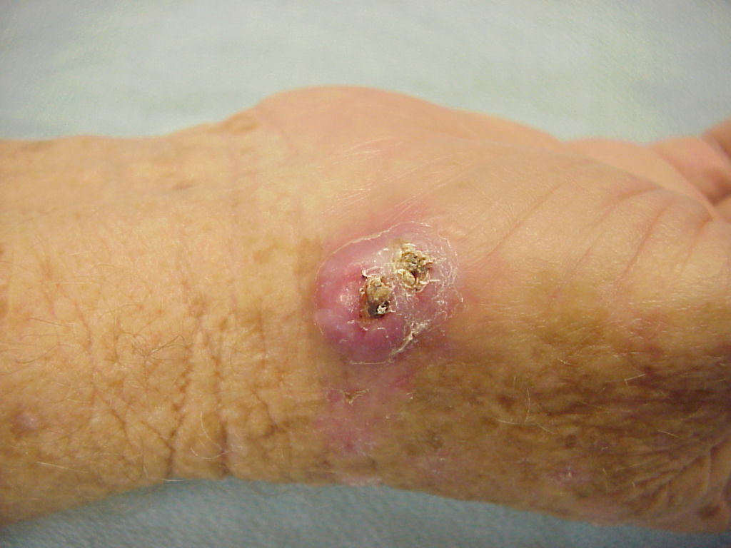
| Keratoacanthoma is a fairly dramatic lesion which most often occurs in sun exposed sites (such as the back of the hand) of a light skinned person. Typically, it appears suddenly and grows rapidly, over the course of weeks. Common clinical features include a central eschar or keratin plug and a raised peripheral border. The usual clinical course is that it grows rapidly, becomes necrotic, and spontaneously regresses over the course of two to four months, leaving a superficial scar. |
| Click on each image for a larger picture |
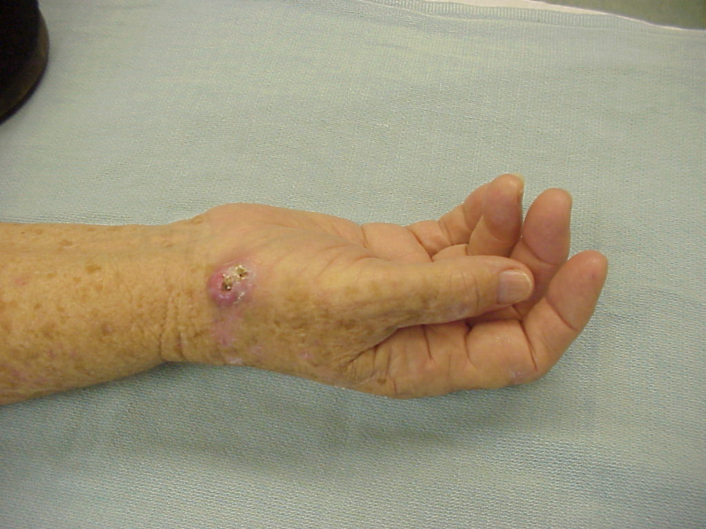
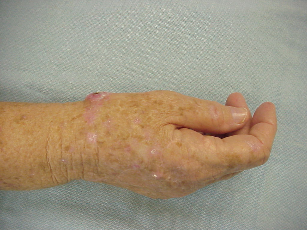
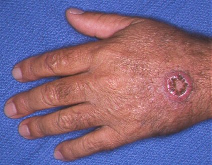
| This rapidly growing crusting lesion was treated with excision and local flap cover. Path - keratoacanthoma. |
 |
 |
 |
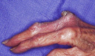
| This resolved spontaneously. |
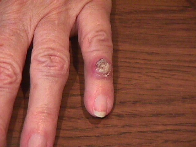 |
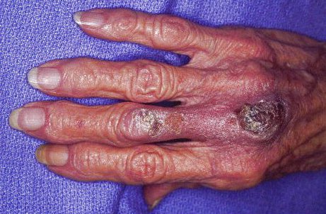
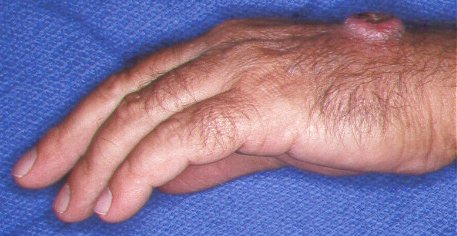
| This crusting skin lesion migrated from the mid dorsum of the proximal phalanx to the metacarpal head area. It was removed with curettage, and healed. Path - keratoacanthoma. |
 |
 |
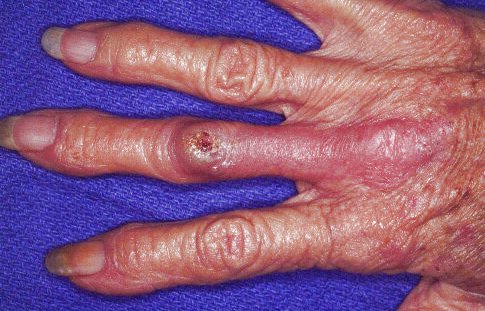 |
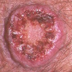
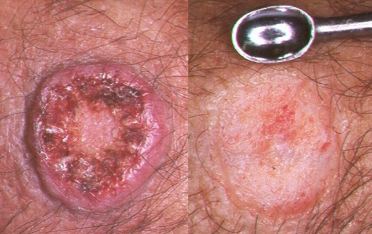
| This keratoacanthoma was treated with curettage. |
 |
 |
 |
| Immediately before and after. The deep dermis is visible at the base of the wound. |
 |
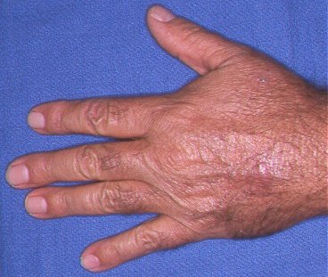
| Late appearance. |
 |
| Search for...
keratoacanthoma |
Case Examples Index Page | e-Hand home |