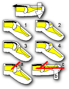
Fig. 17. Mallet finger. The usual injuring forces are active extension and passive flexion of the distal interphalangeal joint. (1.) Soft tissue mallet. (2.) Mallet fracture. (3, 4.) Pediatric Salter I and III mallet fractures. (a,b) Extension block technique for mallet fractures: a pin is placed not through, but just proximal to the fragment to lock it in position. The fracture is then reduced then secured with a second pin extending the distal phalanx